Trichoscopy
Trichoscopy USB Microscope for Hair and Scalp Documentation
Portable magnified viewing for scalp surface, hair shafts, follicular openings, and visual documentation over time.
Overview
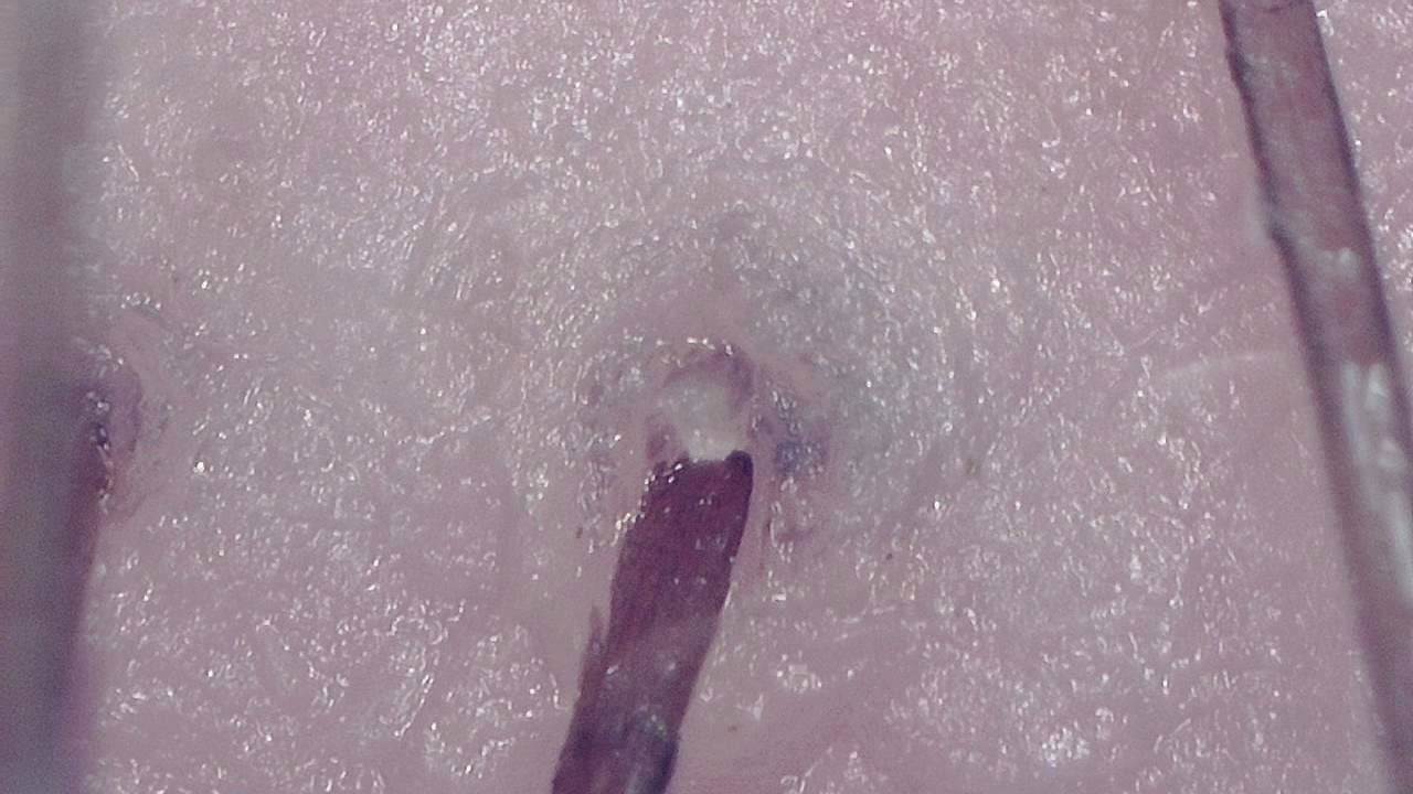
Trichoscopy is a non-invasive visual examination technique used by trained professionals to observe the scalp surface and hair shafts. Smart G-Scope provides a portable USB workflow for magnified viewing and image capture.
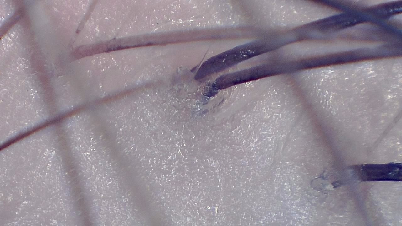
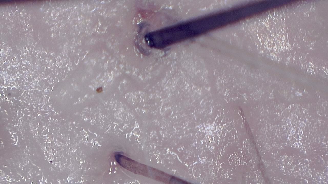
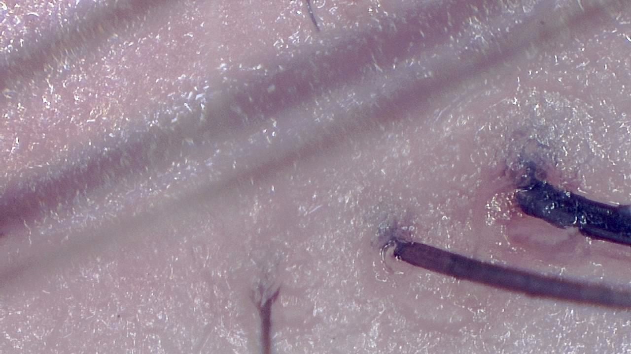
The device can support documentation of hair shaft appearance, scalp surface features, follicular openings, and visible changes over time. It is most useful when images are captured under consistent lighting and positioning.
Smart G-Scope is a visualization and documentation tool. It does not diagnose alopecia or scalp disease by itself; clinical interpretation remains the responsibility of qualified professionals.
Recommended Smart G-Scope setup
- Use a stable hand position or stand to keep the scalp area in focus.
- Clean or part the hair to expose the target scalp area before capture.
- Use consistent magnification, lighting, and labeling for follow-up comparisons.
- Save representative photos or videos through the software for documentation.
What you can observe
- Hair shafts and visible shaft diameter variation.
- Scalp surface texture and visible follicular openings.
- Visible scaling, residues, or surface changes that need professional interpretation.
- Side-by-side image documentation for follow-up visits or patient communication.
Typical workflow
- 1Select and prepare the scalp area to be documented.
- 2Stabilize the microscope and adjust light to avoid glare from hair or skin.
- 3Capture comparable images using consistent distance and magnification.
- 4Label images by area and date so changes can be compared over time.
Key benefits
- Magnified viewing of hair shafts, scalp surface, and visible follicular openings.
- Photo and video capture for documentation and follow-up comparison.
- Portable USB workflow for clinics, hair centers, education, and reports.
- Consistent image records that can support patient communication.
Best for
- Dermatology, trichology, hair clinic, education, and documentation workflows.
- Patient communication where visual records help explain observations.
Not ideal for
- Replacing clinical examination, lab tests, biopsy, or specialist interpretation.
- Hair bulb analysis that requires extraction-based methods.
Image gallery

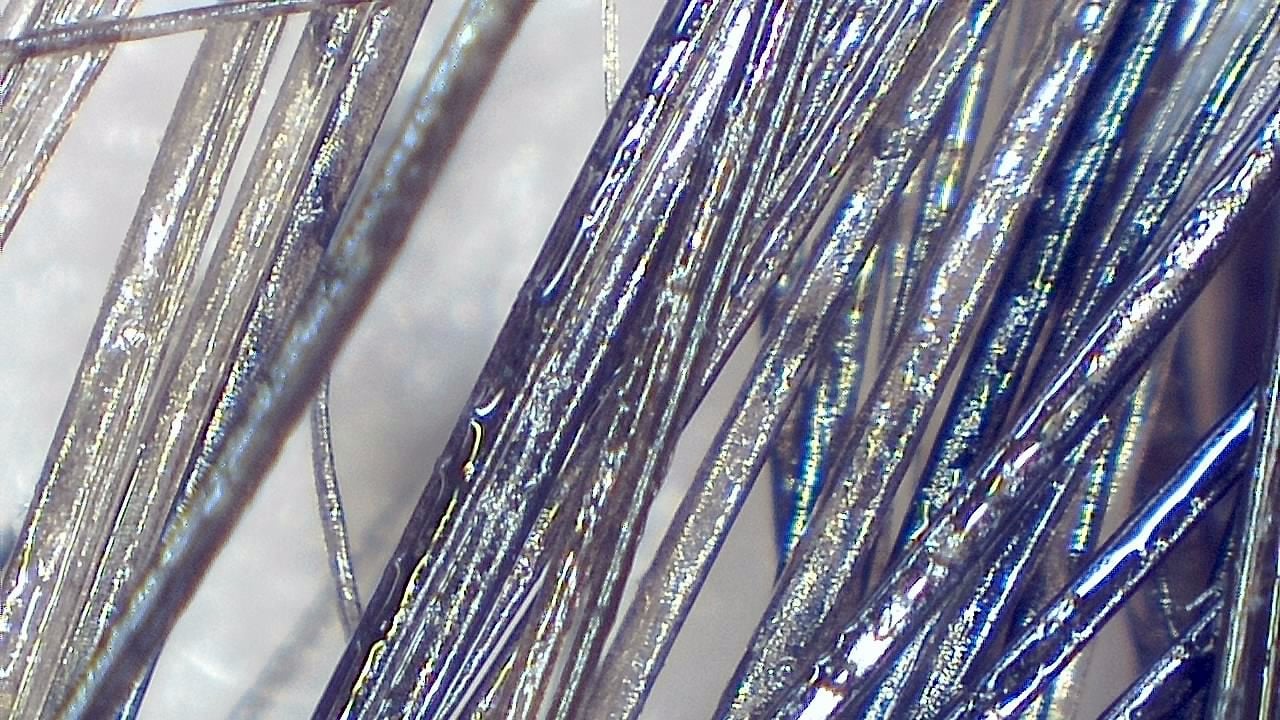
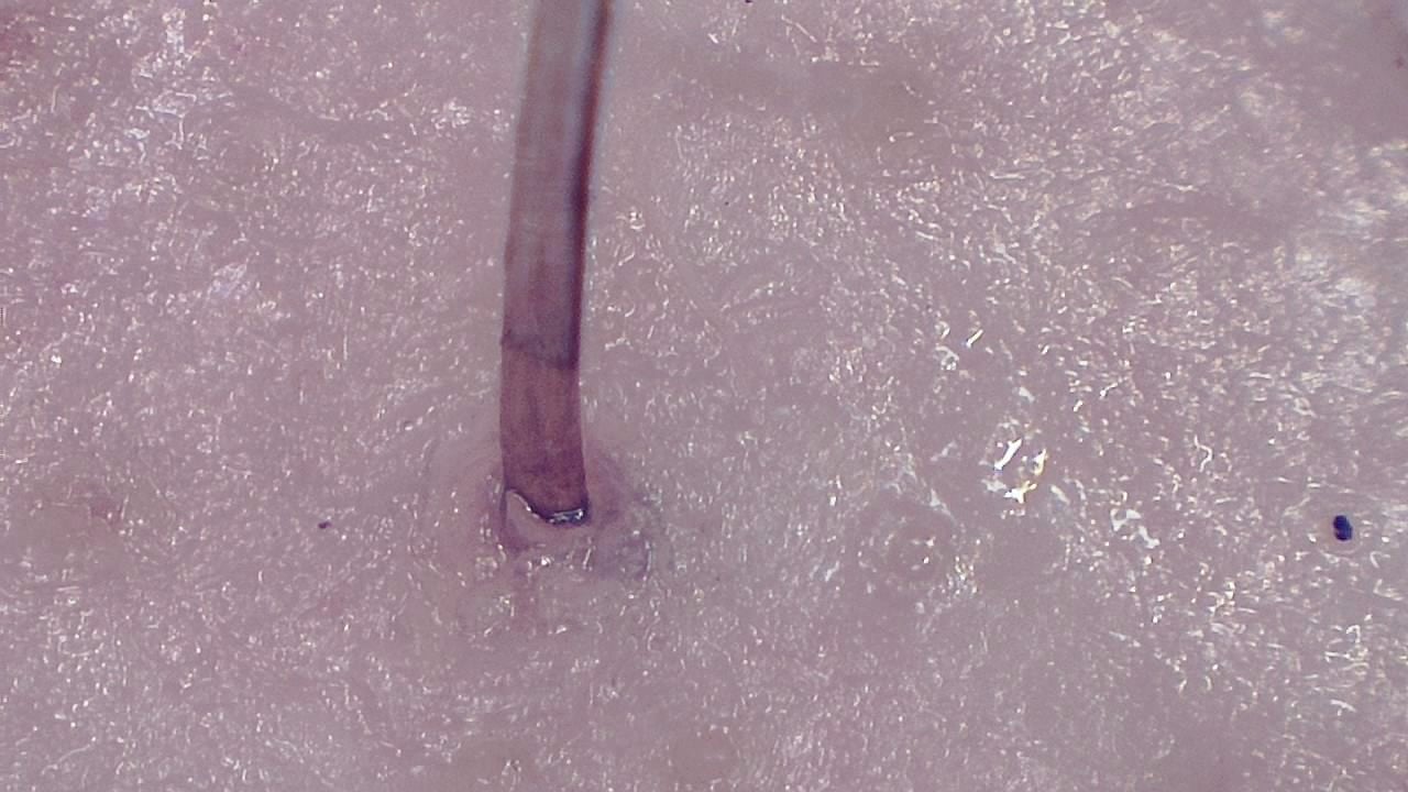
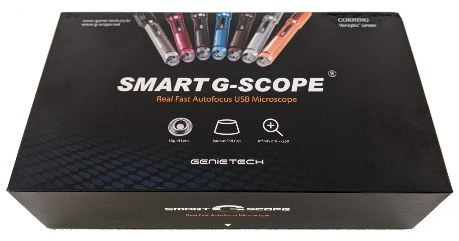
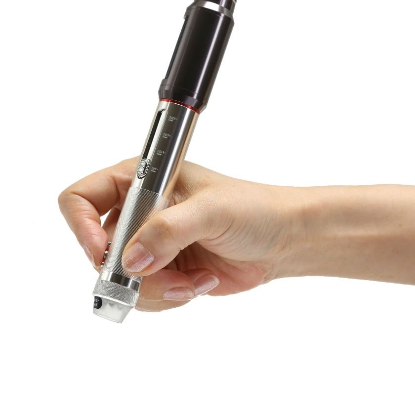
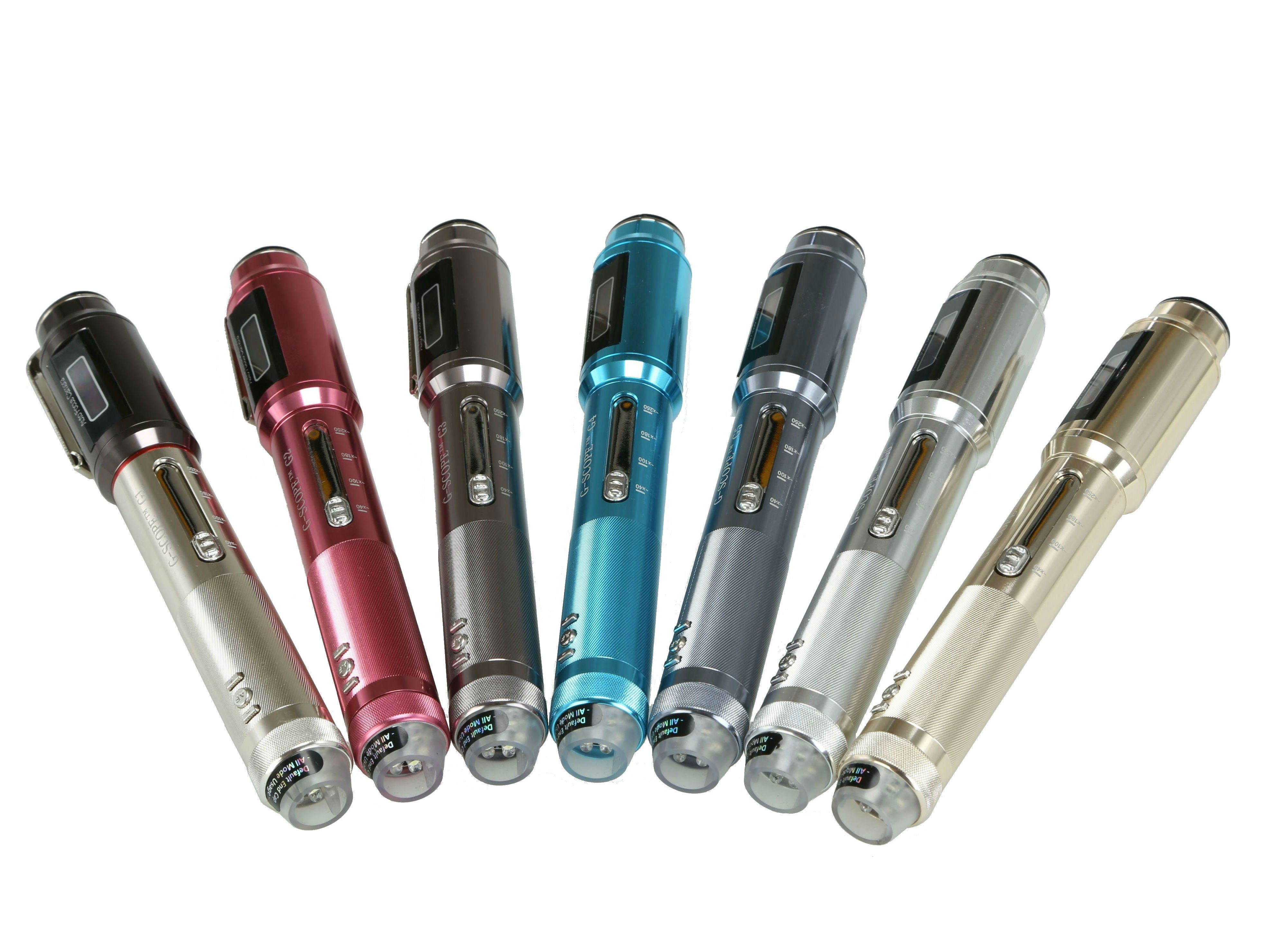
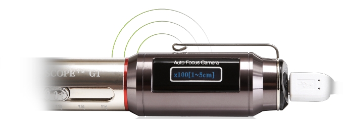
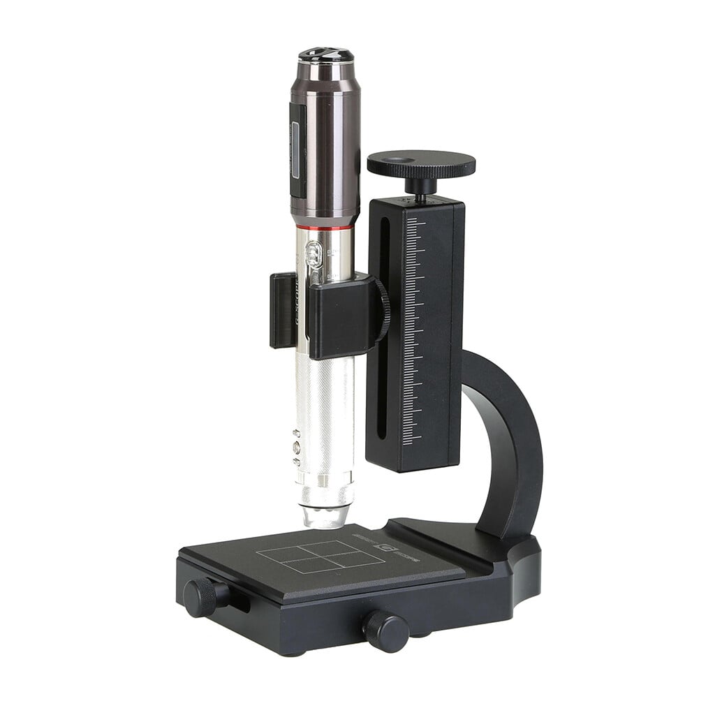
Click any image to enlarge
Practical examples
Baseline scalp documentation
A hair clinic records standardized images from selected scalp areas before follow-up visits, making later visual comparisons easier to discuss.
Post-procedure visual follow-up
After a hair procedure or treatment plan, the team captures comparable images at planned intervals and keeps them with the patient documentation.
Limitations & best practices
- Does not diagnose alopecia or scalp disease by itself.
- Hair bulb analysis requires extraction-based methods outside this workflow.
- Clean or part the hair before capture to avoid cosmetic residues and poor visibility.
- Avoid excessive pressure on sensitive or inflamed areas.
Frequently asked questions
Can Smart G-Scope be used for trichoscopy images?
Yes. It can capture magnified photos or videos of hair and scalp areas for documentation and comparison, provided the area is stabilized and lighting is consistent.
What can I document on the scalp?
Users can document visible hair shafts, scalp surface texture, follicular openings, scaling, residues, and other visible features that require qualified interpretation.
Does Smart G-Scope diagnose hair loss?
No. It supports visualization and documentation. Diagnosis and treatment decisions must be made by qualified professionals.