Dermatology
Clinical Dermatology
High-precision visual analysis of skin lesions, nevi, and dermatologic changes.
Overview
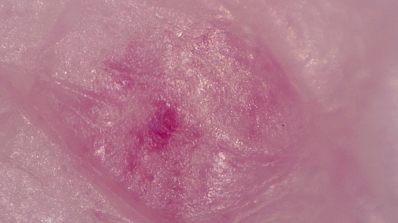
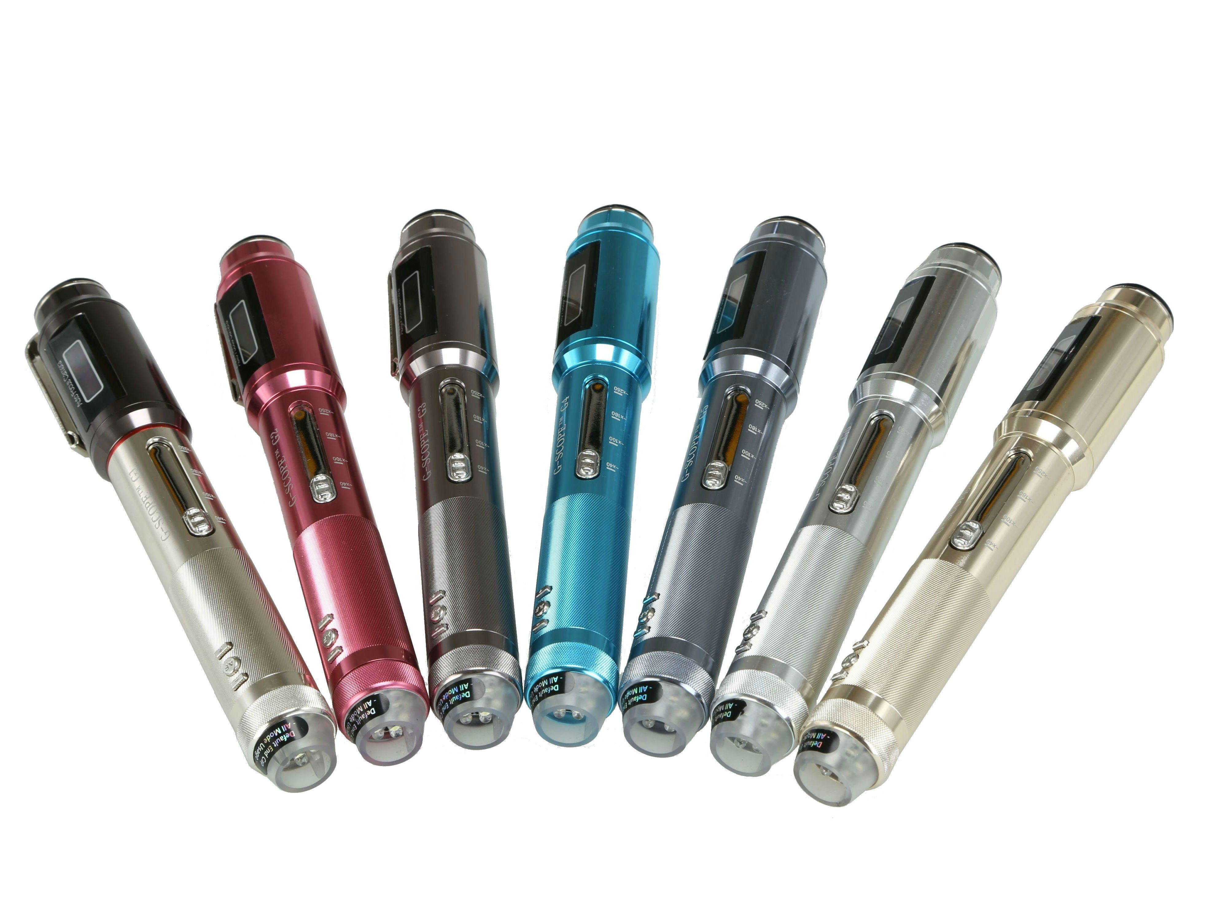
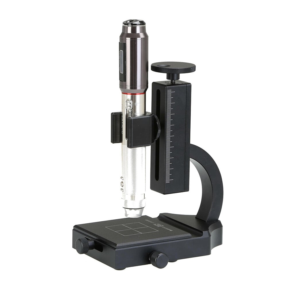
Modern dermatology requires precise visualization tools to document and analyze skin lesions in maximum detail. Smart G-Scope provides dermatology professionals with a portable, high-resolution solution for visual skin examination.
With variable magnification, it is possible to examine everything from superficial structures to deeper epidermal layers, identifying pigmentation patterns, abnormal vascularization, and textural changes that may go unnoticed to the naked eye.
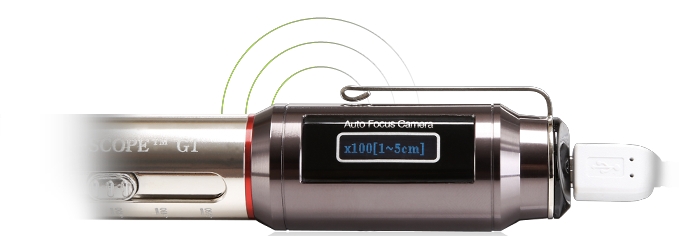
The integrated software enables photo and video capture to support longitudinal lesion follow-up, communication with other specialists, and clinical documentation aligned with healthcare quality standards.
Key benefits
- Visualization of dermal structures with up to 250× magnification
- High-resolution photographic documentation for the clinical record
- Longitudinal lesion follow-up through visual comparisons
- Portability for in-clinic and home visits
Image gallery
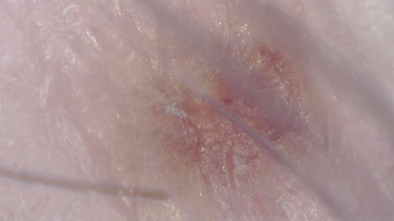
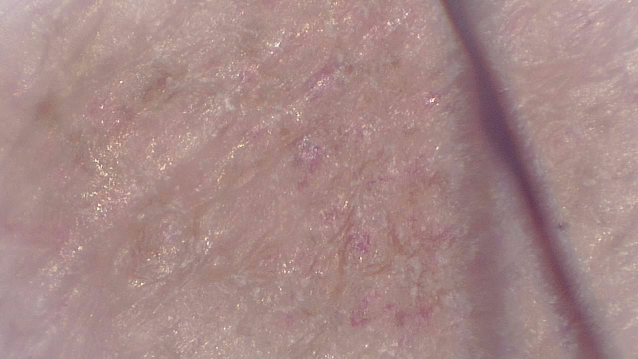
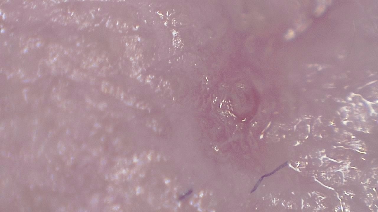
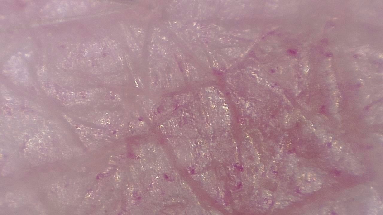
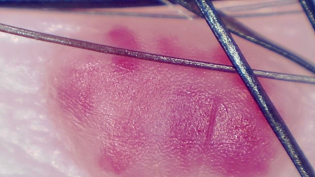
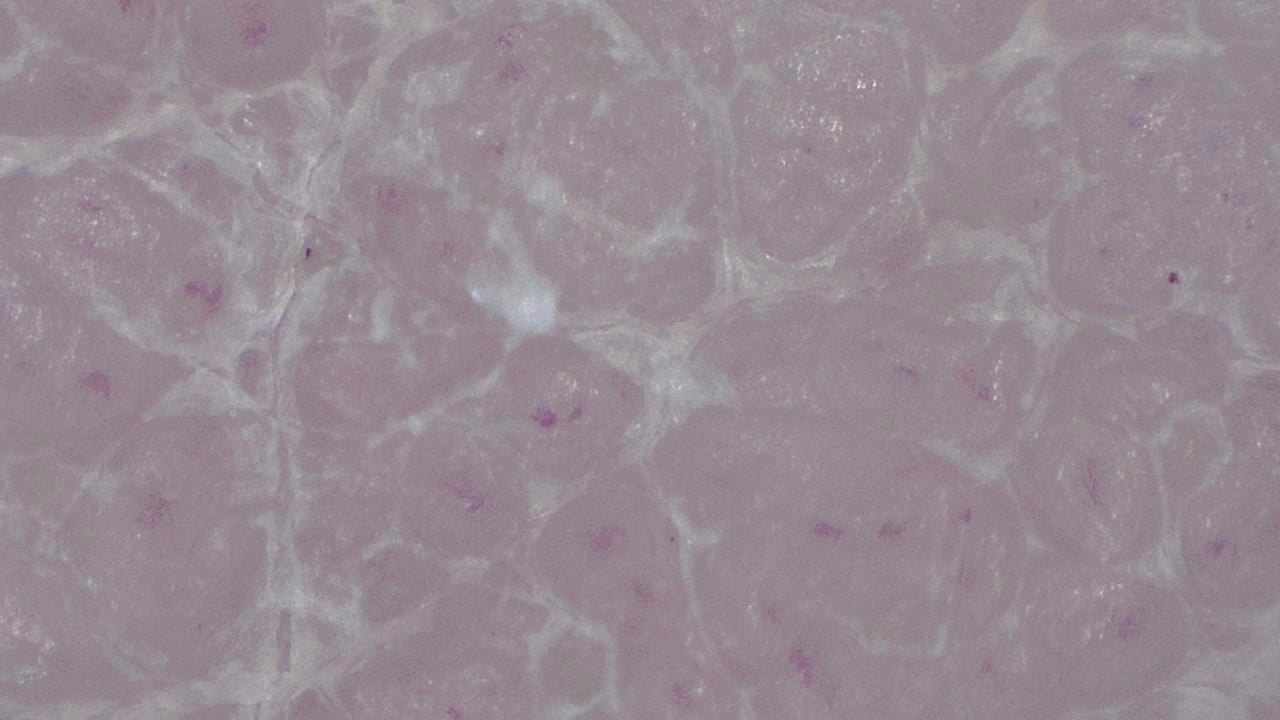
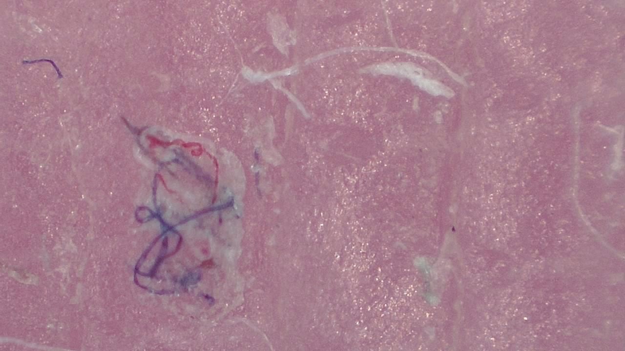
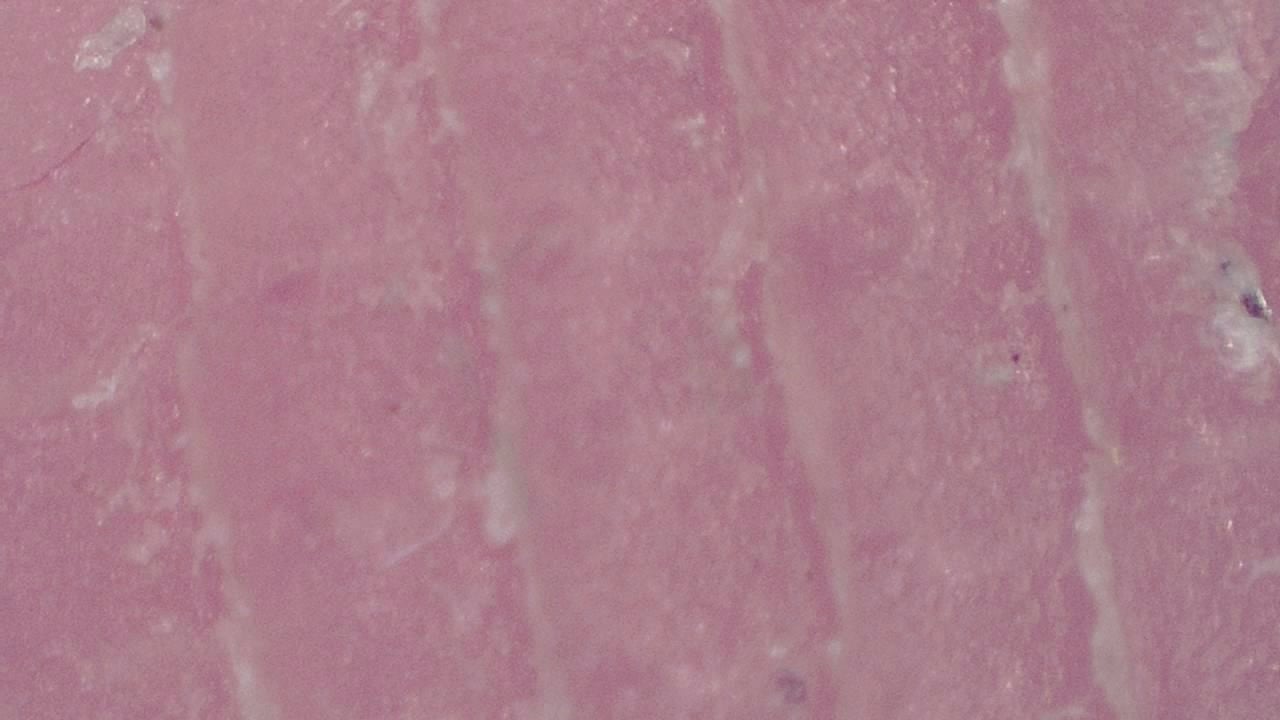
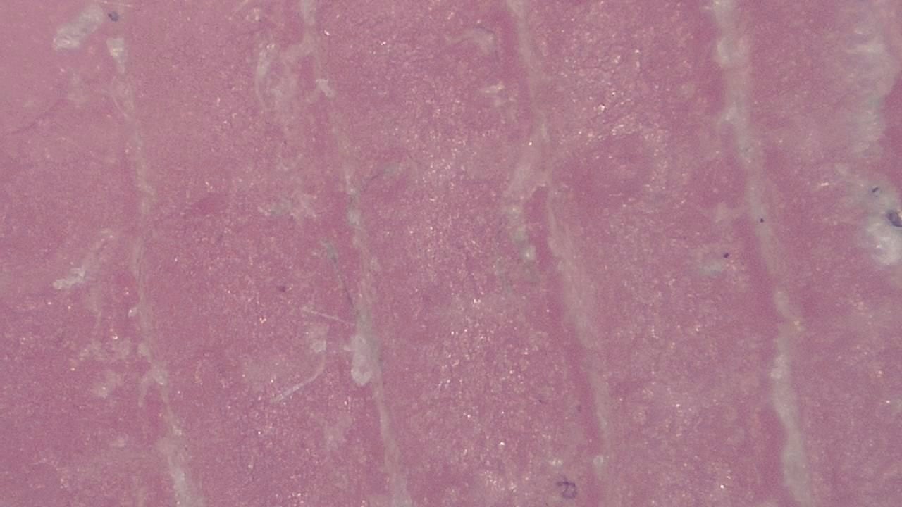
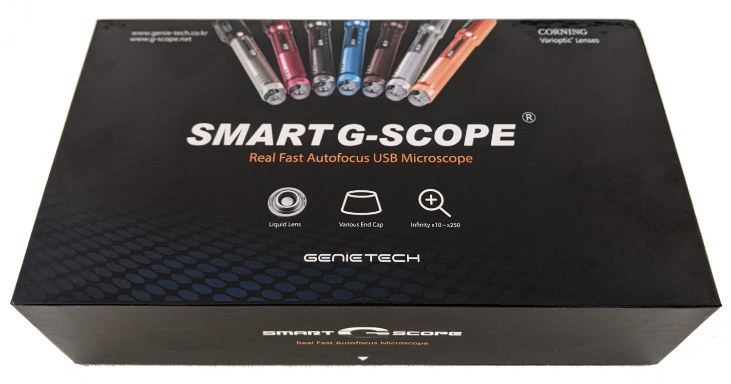
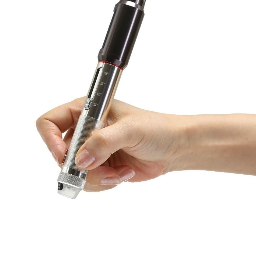
Click any image to enlarge
Practical examples
Atypical nevus follow-up
A dermatologist uses Smart G-Scope to document a suspicious nevus during quarterly check-ups. Comparative images help detect subtle changes in borders and pigmentation, supporting an earlier decision to refer for biopsy before the lesion progresses.
Contact dermatitis assessment
In an allergy clinic, the specialist uses the microscope to analyze the texture and vascularity of eczema-affected areas, documenting treatment response over time and adjusting therapy based on visual findings.
Limitations & best practices
- Images are indicative and do not replace histopathological diagnosis.
- Keep the lens clean to avoid artifacts in captures.
- Adjust illumination according to the patient’s skin phototype.
- Comply with GDPR when storing patient images.