Capillaroscopy
Nailfold Capillaroscopy with Smart G-Scope
A focused workflow for observing nailfold capillaries with the dedicated capillaroscopy head, immersion oil, stable positioning, and image documentation.
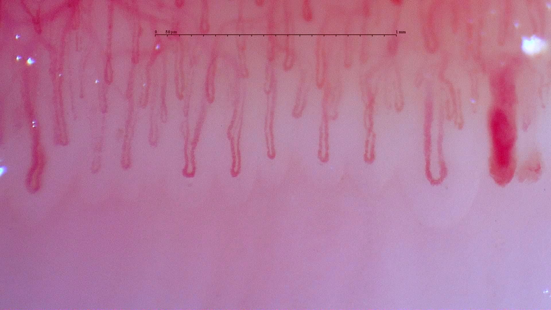
Overview
Nailfold capillaroscopy is used by trained professionals to observe the small capillaries at the nailfold as part of vascular and connective-tissue assessment workflows. The value of the image depends on stable positioning, suitable magnification, controlled light, and consistent capture conditions.
Smart G-Scope supports this workflow when used with the dedicated capillaroscopy head, which is designed for 250x nailfold observation. An immersion medium such as mineral oil or almond oil is used in direct contact with the skin to reduce reflections and improve capillary visibility.
The USB workflow makes it practical to view the image on a computer, capture photos or video, and store visual documentation for follow-up, teaching, referral communication, or distributor demonstrations.
Smart G-Scope Capillaroscope is compatible with Capillary.io CapillaryScope, a desktop application designed for nailfold capillaroscopy using USB capillaroscopes.
Recommended Smart G-Scope setup
- Smart G-Scope with the dedicated capillaroscopy head at 250x.
- Immersion medium in direct contact with the nailfold to reduce reflections.
- A stable hand position or stand to reduce motion blur during capture.
- Computer-based viewing and capture software for photo/video documentation.
Choosing a microscope for capillaroscopy
Magnification and capillary head
Nailfold work requires the dedicated capillaroscopy head at 250x, not just a generic close-up lens.
Stable positioning
Small movements can blur capillary loops, so hand support or a stand matters for repeatable images.
Lighting and immersion
Controlled LED light plus immersion medium reduces reflections and improves nailfold visibility.
Capture and export
A useful setup should make it easy to save images or video for documentation, teaching, or follow-up.
What you can observe
- Nailfold capillary loops and their general arrangement.
- Visible morphology changes such as enlarged capillaries, hemorrhage-like spots, avascular areas, or abnormal branching when present.
- Image sequences that can support documentation and comparison over time.
- Training examples for clinicians, educators, researchers, or distributors.
Typical workflow
- 1Prepare the nailfold area and apply the immersion medium.
- 2Place the capillaroscopy head in controlled contact and stabilize the hand.
- 3Adjust illumination and focus until capillary loops are visible.
- 4Capture representative images or video and label them consistently.
- 5Store or export images according to the clinic, study, or teaching workflow.
Key benefits
- Detailed nailfold capillary visualization at 250x with the dedicated capillaroscopy head.
- Improved visibility when used with an appropriate immersion medium.
- Photo and video capture for documentation, teaching, and follow-up workflows.
- Portable USB setup for clinics, education, research, and distributor demonstrations.
- Compatible workflow with Capillary.io CapillaryScope software.
Best for
- Rheumatology, dermatology, vascular, education, and research teams that need portable nailfold image documentation.
- Distributor demonstrations where a compact USB capillaroscope is easier to show than a large bench system.
- Workflows that need repeatable image capture rather than diagnosis by the device itself.
Not ideal for
- Replacing medical interpretation, serology, biopsy, or other clinical tests.
- Use without the dedicated capillaroscopy head or without a stable contact technique.
Image gallery
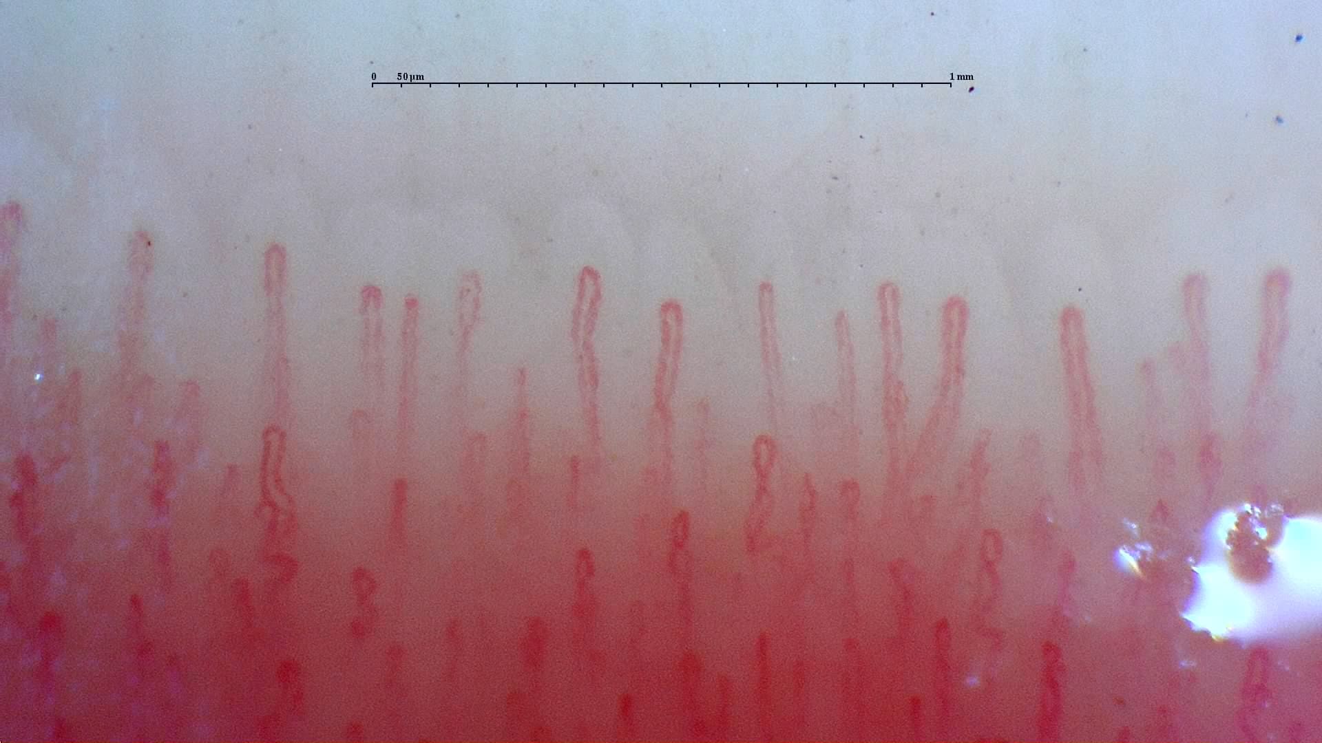
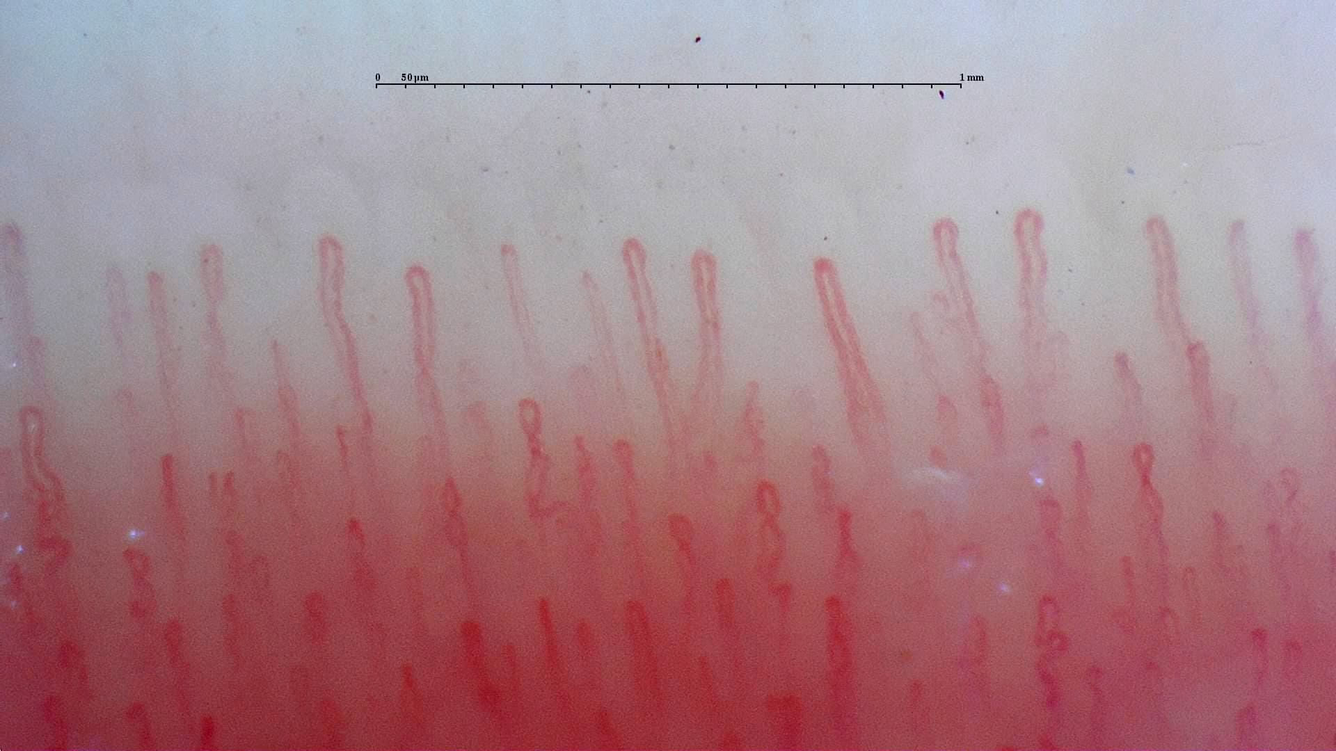
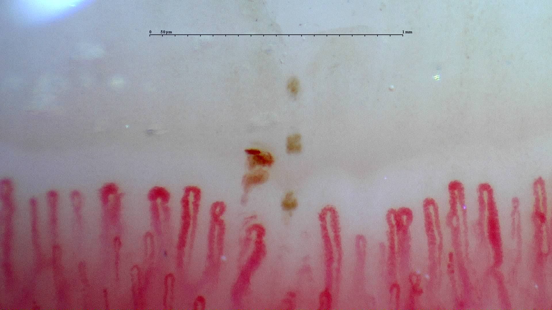
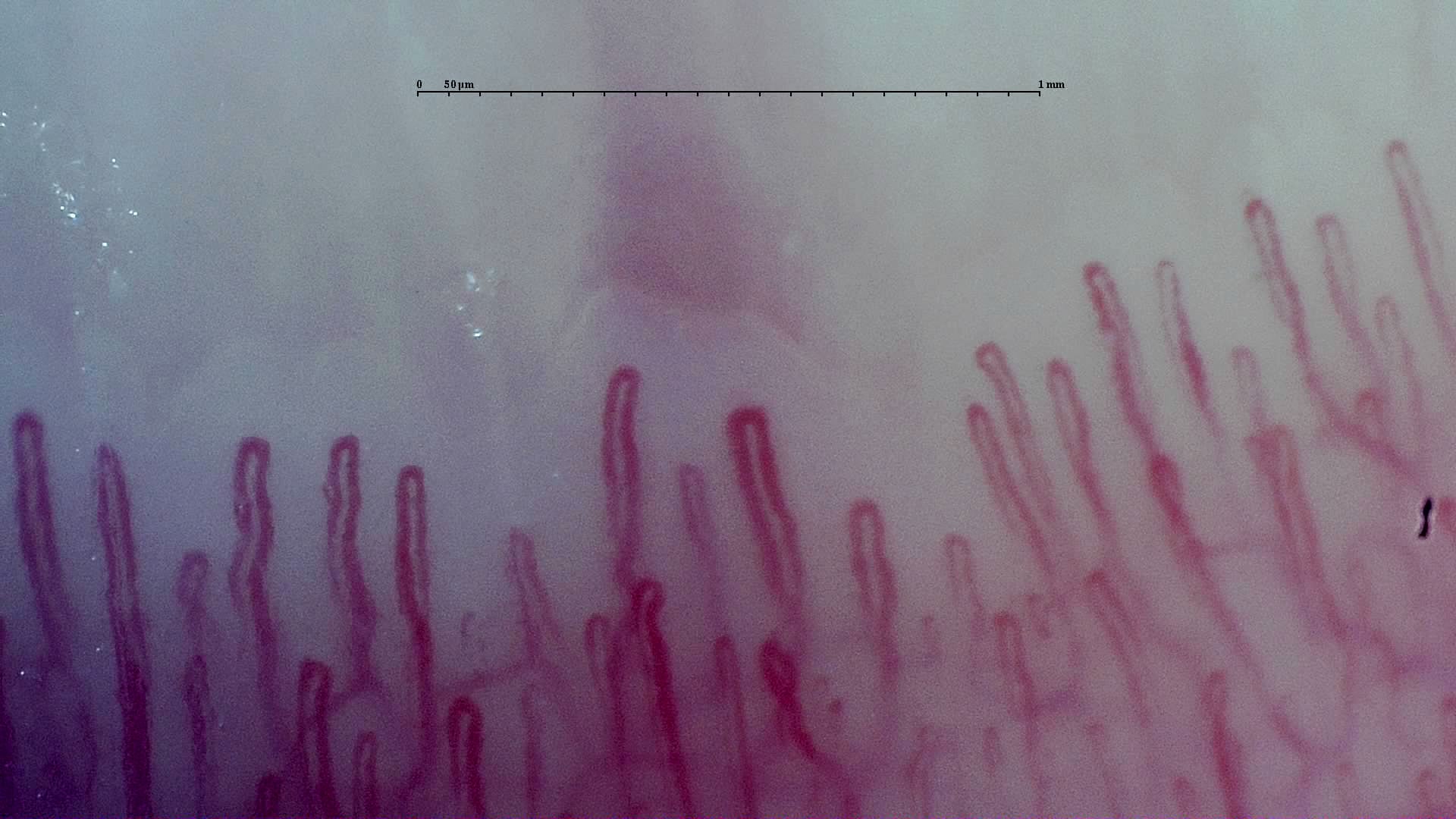
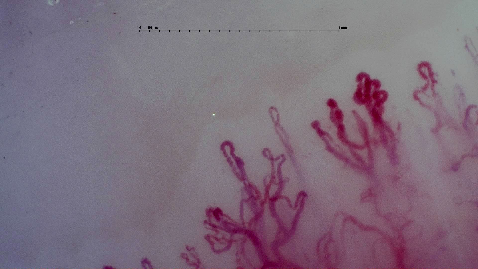
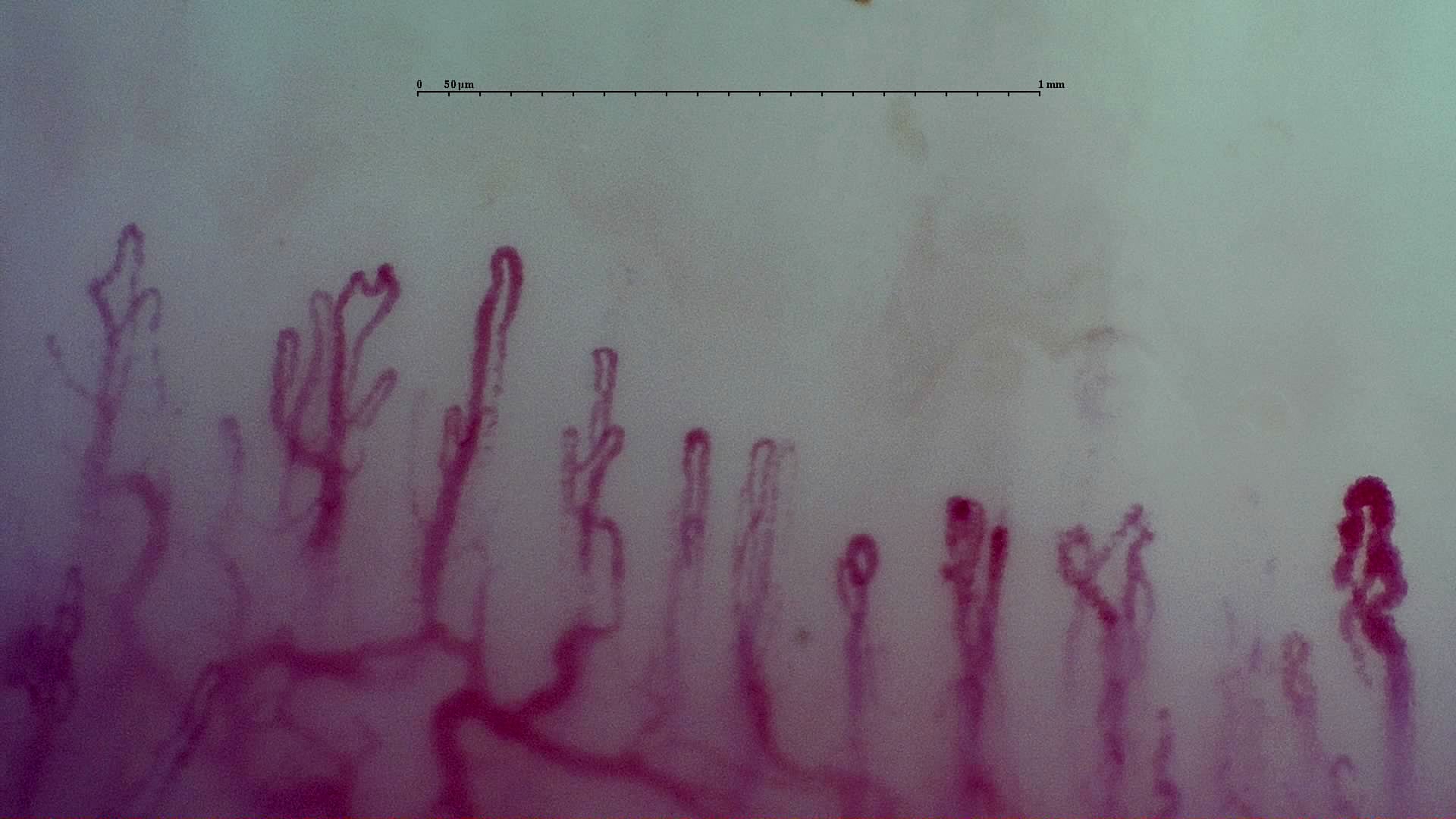
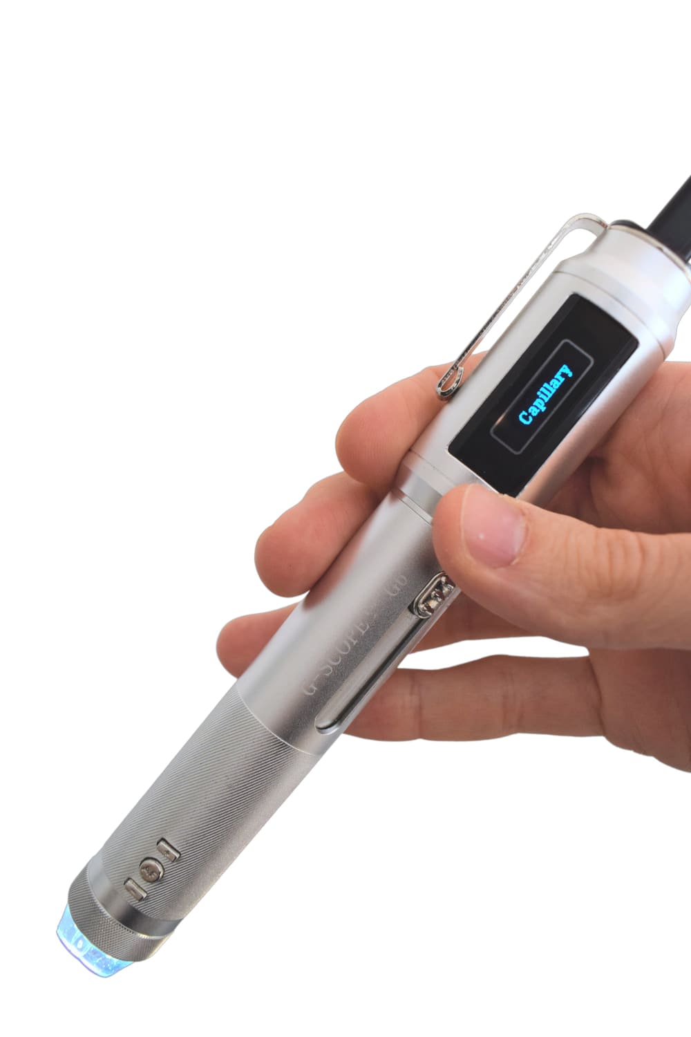
Click any image to enlarge
Practical examples
Raynaud's phenomenon documentation workflow
A clinician captures representative nailfold images with the capillaroscopy head and immersion medium, then stores the images for qualified review and follow-up comparison.
Training and distributor demonstration
An educator or distributor uses the portable USB setup to show how stable positioning, oil contact, and software capture affect nailfold image quality.
Limitations & best practices
- Requires the dedicated capillaroscopy head, functional only at 250x.
- Immersion oil or a comparable immersion medium must be used in contact with the skin.
- Stable positioning is required to reduce motion blur during capture.
- Does not replace medical interpretation, serology, biopsy, or clinical diagnosis.
Frequently asked questions
Is Smart G-Scope suitable for nailfold capillaroscopy?
Yes, when it is configured with the dedicated capillaroscopy head at 250x and used with an appropriate immersion medium and stable positioning. Medical interpretation must be performed by qualified professionals.
What accessories are needed for capillaroscopy?
The dedicated capillaroscopy head is required. A stable stand or controlled hand support is recommended, and immersion oil such as mineral oil or almond oil is used to reduce reflections.
Can images be saved or exported?
Yes. Smart G-Scope workflows support photo and video capture through compatible software so images can be stored for documentation, teaching, or follow-up workflows.
Is Smart G-Scope portable?
Yes. It is a USB microscope, so the setup can be moved between rooms or used with a compatible computer-based workstation.
Is it a replacement for medical diagnosis?
No. Smart G-Scope is a visualization and documentation tool. Diagnosis and clinical decisions remain the responsibility of qualified healthcare professionals.
How do I request pricing or a demo?
Use the contact page and describe your capillaroscopy workflow, preferred software, and whether you need a standalone unit, accessories, or distributor information.